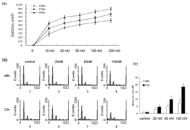
Figure 1.
Bufalin inhibits the proliferation and induces the apoptosis of A549 cells. (a) A549 cells were treated with 10 nmol/L, 20 nmol/L, 50 nmol/L, 100 nmol/L or 200 nmol/L bufalin for 48 h, 72 h, or 96 h. The cell viability was examined by MTT assay. Data were derived from three independent experiments. (b) A549 cells were treated with 20 nmol/L, 40 nmol/L, or 100 nmol/L bufalin for 48 h or 72 h. The apoptosis was examined by PI staining and flow cytometry analysis. (c) Quantification of apoptosis of A549 cells shown in (b). Data were derived from three independent experiments.* P < 0.01 vs. control.

