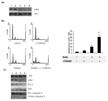
Figure 4.
Bufalin modulates the activation of PI3K/Akt pathway in A549 cells. (a) Western blot analysis of the level of p-Akt in differently treated A549 cells. Total Akt level served as loading control. Shown are representative blots from three independent experiments with similar results: 1. 0 h; 2. 100 nmol/L bufalin for 24 h; 3. 100 nmol/L bufalin for 48 h; 4. 25 μmol/L LY294002 for 2 h plus 100 nmol/L bufalin for 24 h; 5. 25 μmol/L LY294002 for 2 h plus 100 nmol/L bufalin for 48 h. (b) A549 cells were pretreated with 25 μmol/L LY294002 for 2 h followed by treatment with 100 nmol/L bufalin for 48 h as indicated and the apoptosis was examined by PI staining and flow cytometry analysis. Data were derived from three independent experiments. * P < 0.01 vs. left three groups of cells. (c) Western blot analysis of the levels of Bcl-2, Bax, livin and activated caspase-3 in differently treated A549 cells. Total Akt served as loading control. Shown are representative blots from three independent experiments with similar results: 1. 0 h; 2. 100 nmol/L bufalin for 24 h; 3. 25 μmol/L LY294002 for 2 h; 4. 25 μmol/L LY294002 for 2 h plus 100 nmol/L bufalin for 24 h.

