Figure 6.
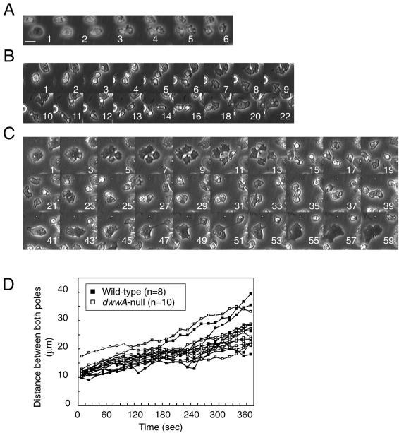
Cytokinetic sequences in wild-type and dwwA-null cells cultured on solid surfaces. Each panel shows a series of phase contrast images recorded at the times indicated at the bottom right (minutes). (A) Mitotic wild-type cells complete cytokinesis within 2-3 min after the onset of furrowing. (B and C) Abortive cytokinesis of mitotic dwwA-null cells. Mitotic dwwA-null cells containing one (B) or two nuclei (C) seemed to have separated completely into two or four daughter cells, respectively, but in fact remained connected by thin cytoplasmic bridges. The connected cells subsequently merged to form a multinucleate cell. Bar, 10 μm. (D) Changes of distances between the poles of two daughter cells of wild-type and dwwA-null cells undergoing cytokinesis. Time zero was chosen when the mitotic cell started to elongate and division axis became apparent. Cells were placed in plastic dishes and images of cell division were analyzed using ImageJ software.

