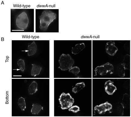
Figure 8.
Localization of myosin II and F-actin in wild-type and dwwA-null cells. (A) Interphase wild-type and dwwA-null cells expressing GFP-myosin II. There is no difference in the distribution of GFP-myosin II in the two cell lines. (B) Cells were fixed with 3.7% formaldehyde and stained with rhodamine-phalloidin for F-actin. In wild-type cells, F-actin was localized in the crowns (arrowheads) and along the cell cortices. The pattern of F-actin distribution in dwwA-null cells differs from that in wild-type cells. Bar, 10 μm.

