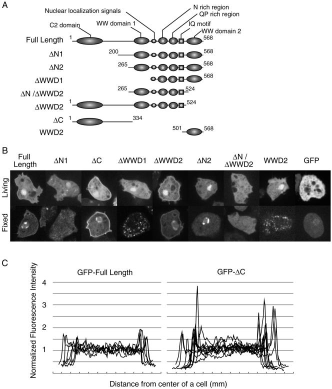
Figure 9.
Localization of a series of DWWA deletion mutants. (A) Schematic representation of the seven constructs. Each box and ellipse shows the indicated domain; the numbers indicate amino acids residues. (B) Fluorescence images of cells expressing GFP-fusion proteins. The top and bottom panels show living cells and cells fixed in 3.7% formaldehyde, respectively. GFP-full length DWWA, GFP-ΔC, and GFP-ΔWWD2 were localized in the cell cortex and nucleus, whereas GFP-ΔN1 and GFP-ΔN/ΔWWD2 were diffusely distributed within the cells, like GFP alone. (C) Profiles of the relative fluorescence intensities of GFP-DWWA and GFP-ΔC in wild-type cells. Fluorescence intensity of the GFP fusion proteins was measured by scanning across the cells, avoiding nuclei, by using NIH Image software. The intensities were then normalized to the average intensity within each cell.

