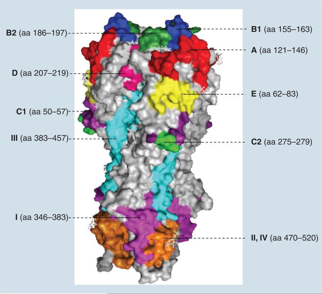
Figure 2. Structure of hemagglutinin and antigenic sites.
The illustration was prepared by using MacPymol© (DeLano Scientific, CA, USA) based on the structure of H3 hemagglutinin (Protein Data Bank accession code 1HGE). The aa forming antigenic site A were marked in red. Antigenic site B1, B2, C1, C2, D and E were marked in blue, forest green, purple, green, pink and yellow, respectively. Antigenic site I in HA2 was marked in magenta, sites II and IV were marked in orange and site III was labeled in cyan.
aa: Amino acid.

