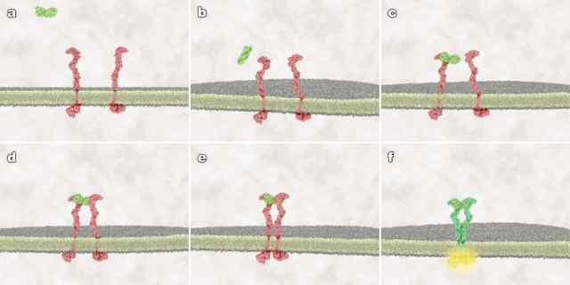
Figure 1.
Static frames from treatment 2 (random protein motion, receptor conformational flexibility, and membrane fluidity) that show the different stages of binding and receptor activation. (A) ligand in the extracellular space, receptor as monomers; (B) ligand approaches membrane, conformationally flexible receptors diffuse in the plane of membrane; (C) ligand randomly encounters and binds a receptor monomer, no receptor activation occurs; (D) ligand-bound and unbound receptors continue to diffuse, trimer forms, no immediate activation occurs; (E) ligand-induced tether of receptor extracellular domains allows cytoplasmic tails to make contact; and (F) contact between cytoplasmic tails leads to cross-phosphorylation and receptor activation.

