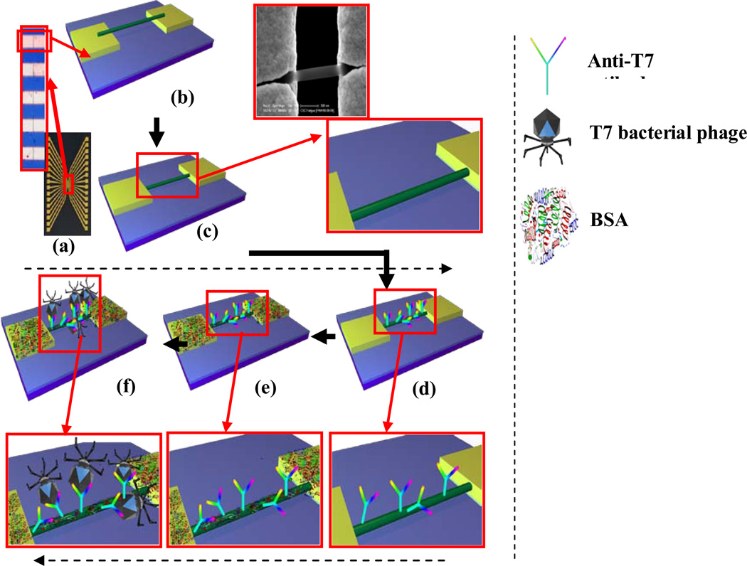
Figure 1.
a) Photo image of 16 electrodes chip (inset optical image showing gap between two electrodes). Schematic of b) aligned and c) anchored Ppy nanowire on the gold electrodes with 3 µm gap (inset: blown up schematic & SEM image). Schematics with blown ups of d) anti-T7 functionalized Ppy nanowire. e) BSA blocking after antibody-functionalization f) T7 phage interacts with anti-T7 antibody on the nanowire surface.

