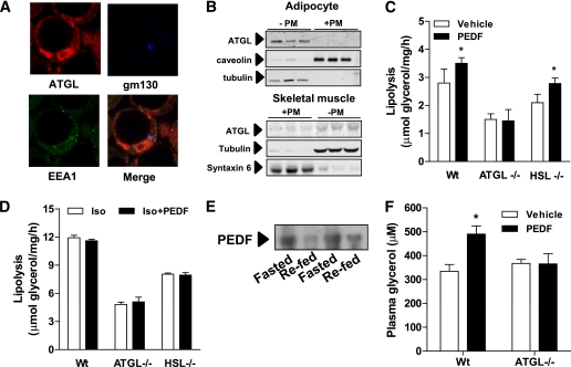
FIG. 1.
ATGL is the major target of PEDF-mediated lipolysis in adipose tissue. A: Immunofluorescence microscopy shows distribution of ATGL in the cytoplasm and around lipid droplets in 3T3-L1 adipocytes. Notably, ATGL is not observed at the plasma membrane (PM) or with endosomal (early endosome antigen 1 [EEA1]: green) or Golgi markers (gm130: blue). B: ATGL colocalizes with the cytoplasmic marker tubulin, but not with membrane markers in a subcellular fractionation. Western blot shows location of various subcellular markers in a fractionation from 3T3-L1 adipocytes (top) and L6 myotubes (below). +PM, plasma membrane–containing fraction. C: Epididymal fat pads were excised from lean ATGL−/−, HSL−/−, or Wt littermates, and lipolysis was assessed as glycerol release into the buffer. PEDF: 100 nmol/L (n = 4 per group). *P < 0.05 vs. vehicle within the same genotype. D: PEDF (100 nmol/L) does not affect β-adrenergic–stimulated lipolysis in epididymal fat pads. Fat pads were incubated for 2 h in isoproterenol (1 μmol/L), and glycerol release was determined (n = 4 per group). E: Immunoblot of plasma PEDF in mice fasted for 16 h or 2 h after refeeding. F: In vivo lipolysis is increased by PEDF. Lean ATGL−/− or Wt mice were injected with PEDF intraperitoneally, blood was obtained after 30 min, and glycerol was assessed in the plasma (n = 6 mice per group). *P < 0.05 vs. vehicle. Data in graphs are mean ± SEM. (A high-quality digital representation of this figure is available in the online issue.)

