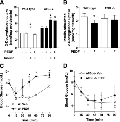
FIG. 5.
PEDF causes skeletal muscle insulin resistance in an ATGL-dependent manner. A: 2-Deoxy-d-glucose (2-DG) uptake experiments in primary myotubes. Wt (□) and ATGL−/− (■) myotubes were pretreated with saline or PEDF (100 nmol/L) for 2 h. The media was removed, and basal and insulin-stimulated 2-DG uptake was determined (n = 6 for each group where each experiment was performed in triplicate on two occasions). *P < 0.05 vs. vehicle within the same genotype. Values are means ± SEM. B: Extensor digitorum longus muscles were removed from ATGL−/− (■) and Wt littermates (□) before assessment of insulin-stimulated glucose uptake (n = 4–6 for each group where each experiment was performed on two occasions). *P < 0.05 vs. –PEDF within the same genotype. Values are means ± SEM. Insulin tolerance tests were performed in Wt (C) and ATGL−/− mice (D). PEDF was injected intraperitoneally 2 h before insulin administration. PEDF (n = 6–7 per treatment). *P < 0.05 vs. corresponding time point within the same genotype.

