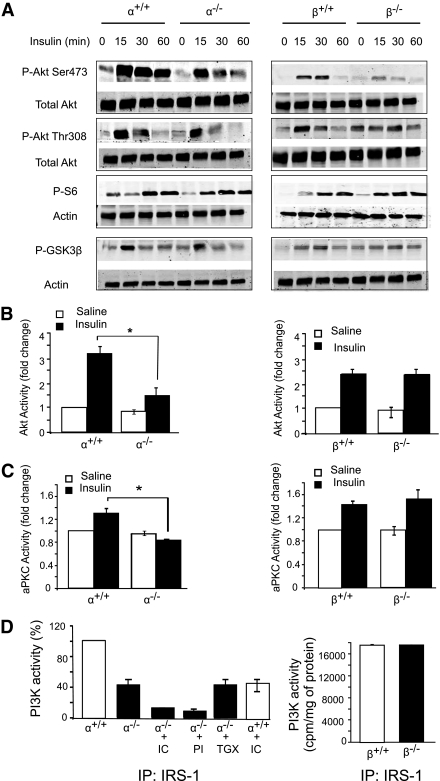
FIG. 2.
Insulin signaling in p110-null livers. A: Liver lysates from fasted mice treated intraperitoneally without or with insulin (2 units/kg body wt) for the indicated times were analyzed by Western blotting. B and C: Fasted mice were injected intraperitoneally with saline or insulin (2 units/kg body wt). Livers were collected 15 min later, and lysates were prepared and assayed for Akt (B) or aPKC (C) kinase activity. *P < 0.05 by t test (N = 3 for all groups). D: Mice were fasted overnight and then injected through the inferior vena cava with insulin (2 units/kg body wt). Livers were collected 5 min later, and lysates were subjected to immunoprecipitation with IRS-1 antibody. The immunoprecipitates for p110-α+/+ and p110-α−/− (left panel) were assayed for PI3K activity in the presence of vehicle, 500 nmol/L PI-103 (PI), 50 nmol/L TGX-221 (TGX), or 5 μmol/L IC87114 (IC). Values are normalized to the average PI3K activity in p110-α+/+ samples assayed in the absence of inhibitors. The immunoprecipitates for p110-β+/+ and p110-β−/− (right panel) were assayed without any additions (N = 3 for all groups). Values shown are mean ± SEM.

