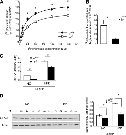
FIG. 5.
Loss of p110-α attenuates fatty acid uptake in hepatocytes. Hepatocytes were isolated from p110-α+/+ and p110-α−/− mice fed normal chow as described in research design and methods. A: Cells were incubated with increasing amounts of [3H]palmitate/BSA at a 3:1 molar ratio for 1 min. [3H]palmitate uptake into the cell was then measured. *P < 0.001 by t test. B: Hepatocytes were incubated with [3H]palmitate/BSA for 2 h, and [3H]palmitate incorporated into cellular triglycerides was then measured. *P < 0.001 by t test. C: Quantitative RT-PCR analysis of mRNA levels in livers of p110-α+/+ and p110-α−/− mice fed an HFD or normal chow (NC). *P < 0.05 by t test (N = 6). D: Liver lysates were analyzed by Western blotting with the indicated antibodies. Bands were quantified using densitometry. *P < 0.005 by t test. Values are means ± SEM of three independent experiments done in triplicate.

