FIG. 3.
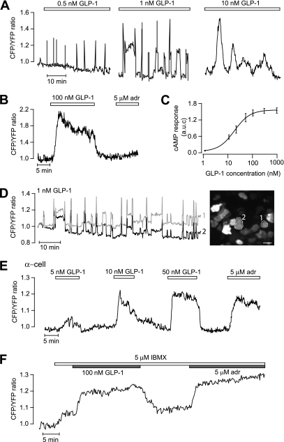
GLP-1 triggers [cAMP]pm oscillations that are synchronized between islet β-cells. TIRF microscopy recordings of [cAMP]pm in individual cells within an intact mouse pancreatic islet exposed to 3 mmol/L glucose. A: [cAMP]pm oscillations in a β-cell exposed to increasing concentrations of GLP-1 (the trace was interrupted between changes in GLP-1 concentration). The amplitude and duration of individual oscillations tended to increase with the GLP-1 concentration. B: Stable and reversible [cAMP]pm elevation in response to 100 nmol/L GLP-1 in a β-cell identified by the lack of adrenaline (adr) effect. C: Dose dependence of the GLP-1 induced time-average [cAMP]pm elevation in islet β-cells. Means ± SEM for 4–9 cells at each concentration. D: [cAMP]pm oscillations induced by 1 nmol/L GLP-1 are synchronized between β-cells lacking apparent direct contact. The recordings are from the two cells in the TIRF image encircled with broken lines. Scale bar, 10 μm. E: Example of a rare α-cell with dose-dependent [cAMP]pm elevations in response to GLP-1. F: GLP-1 response in an adrenaline-identified α-cell exposed to 5 μmol/L IBMX. (A high-quality digital representation of this figure is available in the online issue.)

