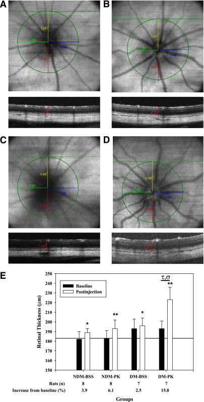
FIG. 7.
Representative en face OCT and B-scan images of rat retina obtained by spectral domain optical coherence tomography (SD-OCT) at 24 h after intravitreal injection and resultant retinal thickness measurements. After 4 weeks’ diabetes duration, baseline SD-OCT scans of the retina from each eye were obtained (100 B-scans of 1,000 A-scans over a 1.5 × 1.5 mm area). Rats were given 10-μL intravitreal injections of PK (50 ng/eye) and BSS in the contralateral eye. At 24 h after injection, repeat SD-OCT scans of the retina from each eye were obtained. A–D: The center of the optic nerve was identified, and a 500-μm radius was drawn on the en face image as shown in. The intersection of the radii terminus and the corresponding B-scan identified the retinal sites for thickness measurement. Retinal thickness was measured on the intersecting B-scan from the RPE to the ILM using calipers calibrated in microns. En face images were saved for retinal registration between timed SD-OCT scans. En face and B-scan slices are shown for nondiabetes (NDM) plus BSS (A), nondiabetes plus PK (B), diabetes (DM) plus BSS (C), and diabetes plus PK (D). B-scans correspond to the horizontal green line shown on each en face image. A single caliper is drawn on the corresponding B-scan at the 500-μm point from the ONH. B-scans of PK-treated nondiabetic and diabetic retina illustrate increased thickness compared with BSS alone. En face images show vessel tortuosity and vascular dilation of the primary vasculature after PK injection compared with BSS-treated retina. E: Quantification of retinal thickness in diabetic and age-matched nondiabetic rats at baseline and 24 h after intravitreal injection of PK. Diabetes was induced in rats by STZ injection. After 4 weeks of diabetes, baseline SD-OCT scans of the retina from each eye were obtained. Rats were given 10-μL intravitreal injections of PK (50 ng/eye) and BSS in the contralateral eye. At 24 h postinjection, follow-up SD-OCT scans of the retina from each eye were obtained. For BSS-treated eyes, retinal thickness increased at 24 h by 3.9% (*P < 0.05) and 2.5% (*P < 0.05) compared with baseline thickness in nondiabetic and diabetic rats, respectively. For PK-treated eyes, retinal thickness increased at 24 h by 6.1% (**P < 0.01) and 15.8% (P < 0.01) compared with baseline thickness in nondiabetic and diabetic rats, respectively. The increase in retinal thickness by PK in diabetes was greater than the change observed in the contralateral BSS injection alone (τP = 0.002) and was greater than the effect of PK on thickness in nondiabetes (σP = 0.017). (A high-quality digital representation of this figure is available in the online issue.)

