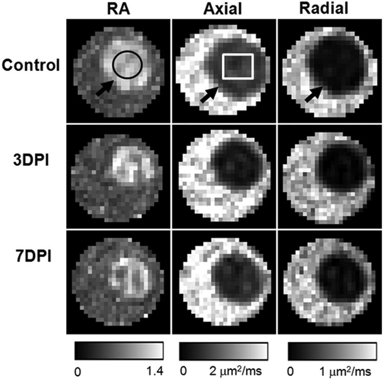
Figure 1.

DTI-derived RA, axial diffusivity, and radial diffusivity of control (top row), 3 days postischemia (DPI) (middle row), and 7DPI (bottom row) experiment groups. The optic nerve (black arrows) was embedded in a tube filled with PBS. The optic nerve was readily differentiated from the PBS in all DTI parameter maps, which simplified region of interest (ROI) placement. The black circle is the ROI for data analysis. The white rectangle signifies the region for quantitative immunohistochemistry staining.
