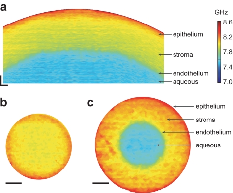
Figure 2.
Brillouin imaging of the cornea. (a) A cross-sectional Brillouin image of bovine cornea, revealing the decreasing modulus with depth. The horizontal (x) and vertical (z) span is 5 × 0.5 mm. (b) En face Brillouin image of the cornea optically sectioned at a shallow depth. (c) A Brillouin image of a deeper section. Scale bars: (a) 200 μm; (b, c) 1 mm.

