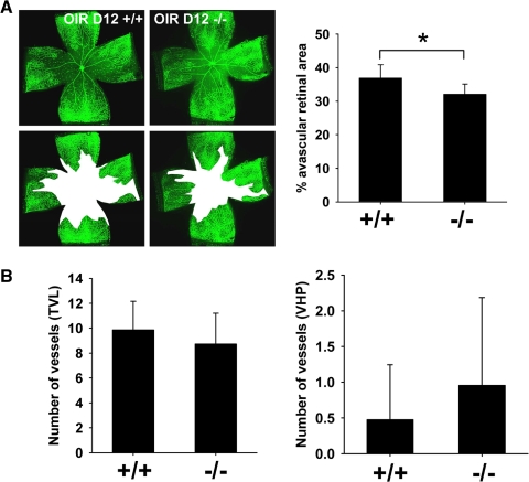
Figure 4.
Optc−/− mice display a decrease in vaso-obliteration in the oxygen-induced retinopathy model at P12. (A) Optc+/+ and Optc−/− animals were subjected to the OIR model and euthanized at P12. The central avascular area was outlined in white after fluorescent isolectin staining at OIR P12 and quantification was performed as a percentage of the total retinal area. This revealed a 13% decrease in vaso-obliteration in Optc−/− (n = 10) compared with Optc+/+ (n = 7) mice and this was statistically significant (*P = 0.012). (B) Quantification of the number of vessels in TVL and VHP after H&E staining of eye sections at this stage showed no significant difference between Optc+/+ (n = 7) and Optc−/− (n = 10) (P = 0.348 for TVL and P = 0.390 for VHP).

