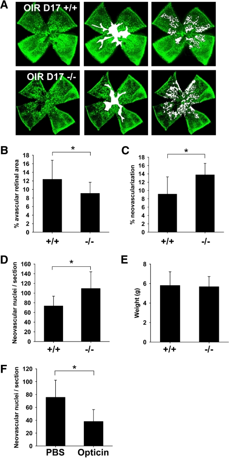
Figure 5.
Optc−/− mice display an increase in preretinal neovascularization in the oxygen-induced retinopathy model at P17. (A) Optc+/+ and Optc−/− mice were subjected to the OIR model and then analyzed at P17. Quantification of vaso-obliteration (outlined in white in middle panel) and preretinal neovascularization (highlighted with white dots in right panel) was performed on retinal flat-mounts after fluorescent isolectin staining (using SWIFT_NV software) and reported as a percentage of total retinal area. (B) There was a significantly less (26%) vaso-obliteration in the Optc−/− mice (n = 37/from 3 litters) compared with the Optc+/+ counterparts (n = 20/from 3 litters) (*P = 0.006). (C) The Optc−/− mice (n = 37/from 3 litters) demonstrated 51% more preretinal neovascularization than the Optc+/+ mice (n = 20/from 3 litters) (*P < 0.001). (D) The neovascular response at OIR P17 was also quantified by counting ECs in preretinal vascular tufts on H&E-stained eye sections and revealed that Optc−/− mice (n = 8/from 2 litters) developed 46% more preretinal ECs than Optc+/+ mice (n = 8/from 3 litters) (*P < 0.001). (E) The weight of Optc+/+ (n = 15) and Optc−/− (n = 26) mice was measured at P17 after being subjected to the OIR model and there was no significant difference between both animals (P = 0.746). (F) In further experiments, one eye from Optc+/+ mice subjected to the OIR model was injected with recombinant opticin (n = 6/from 2 litters) at P14 and the contralateral eye with PBS (carrier buffer), then neovascular growth analyzed at P17 by counting ECs in preretinal vascular tufts on H&E-stained eye sections. Quantification of neovascularization revealed that eyes injected with opticin had 50% less preretinal ECs than PBS-injected control eyes (*P < 0.001). Similar opticin injection experiments (one eye receiving opticin and the contralateral eye buffer alone) were performed with Optc+/+ eyes (n = 6/from 1 litter), but quantification of vaso-obliteration and preretinal neovascularization was performed on retinal flat-mounts after fluorescent isolectin staining (SWIFT_NV software). No significant difference could be observed between both groups in the hyperoxic-driven vaso-obliterative response (P = 0.960), but the opticin injected Optc+/+ eyes developed significantly less preretinal neovascularization compared with PBS control (P < 0.001) (data not shown).

