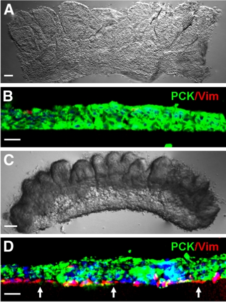
Figure 1.
Collagenase but not dispase isolates more subjacent Vim+ cells. Dispase digestion of the one-clock-hour limbal segment at 4°C for 16 hours removed the entire PCK+ epithelial sheet (A), which consisted predominantly of PCK+ (green) cells (B). In contrast, collagenase digestion at 37°C for 18 hours isolated a cluster (C), which contained significantly more closely associated PCK-/Vim+ (red) cells (D, arrows). Nuclear counterstaining by Hoechst 33342. Scale bars: 100 μm (A, C); 20 μm (B, D).

