Figure 3.
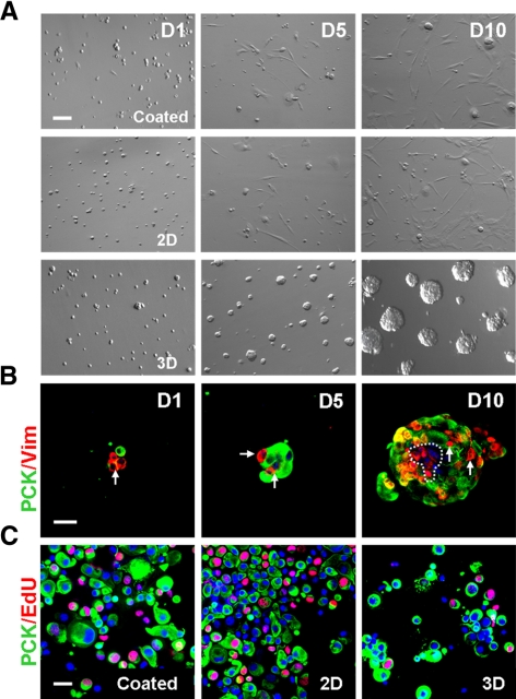
Different growth by collagenase-isolated cells in coated, 2D, and 3D Matrigel. Single cells from collagenase-isolated limbal clusters (Fig. 1) were seeded in coated, 2D, and 3D Matrigel at 5 × 10⁴/cm2 in MESCM. Spheres emerged in 3D Matrigel, whereas predominant spindle cells were found in coated and 2D Matrigel (A). The sphere in 3D Matrigel was formed by the reunion of single PCK+ (green) cells and Vim+ (red) cells, both of which increased in cell numbers in 10 days (B). Double staining between PCK (green) and EdU (red) confirmed that EdU+ nuclei were high in coated and 2D Matrigel but low in 3D Matrigel (C). Scale bar, 20 μm.

