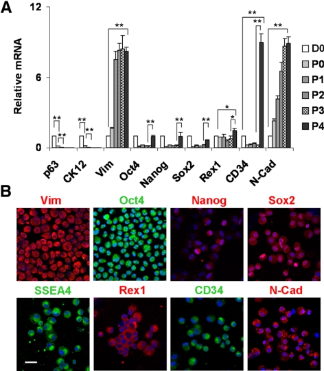
Figure 5.
Phenotypic characterization of expanded mesenchymal cells. Compared with D0 clusters immediately isolated by collagenase, qRT-PCR revealed the rapid disappearance of p63 and CK12 transcripts by P2, a significant decline of Oct4, Nanog, Sox2, and CD34 (n = 3, **P < 0.01), a steady increase of Vim and N-Cad (n = 3, **P < 0.01), and no change in Rex1 from on coated Matrigel from P0 to P3 (A). On reseeding in 3D Matrigel at P4, Oct4, Nanog, Sox2, Rex1, and CD34 transcripts were significantly increased (n = 3, *P < 0.05 and **P < 0.01, compared with P3 cells) (A). All cells derived from P4 aggregates were Vim+ and heterogeneously expressed SC markers (B). Scale bar, 20 μm.

