Figure 8.
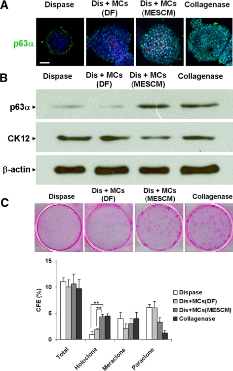
Maintenance of limbal epithelial progenitor status by MCs Expanded in MESCM but not DF. D10 spheres in 3D Matrigel were formed by dispase-isolated limbal epithelial cells alone (dispase) or were mixed with MCs expanded on coated Matrigel in DF (Dis+MCs [DF]) or in MESCM (Dis+MCs [MESCM]) or by collagenase-isolated clusters (Collagenase). Immunofluorescence staining of p63α demonstrated that Dis+MCs (MESCM) had more p63α (green) expression than Dis+MCs (DF) (these MCs were prelabeled by nanocrystals [red]) (A). Western blot analysis confirmed that dispase+MCs (MESCM) had more p63α but less CK12 than dispase+MCs (DF) using β-actin as the loading control (B). Spheres generated by Dis+MCs (MESCM) had significantly more holoclone than those by Dis+MCs (DF) using dispase and collagenase as controls (C, n = 3, **P < 0.01). Scale bar, 20 μm.

