Figure 5.
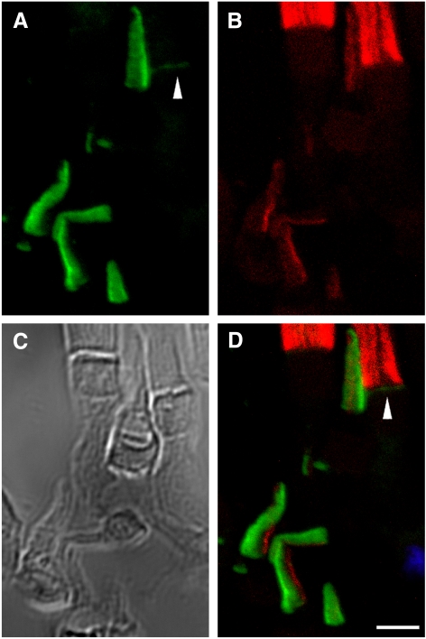
Double immunolabeling of X. laevis photoreceptors with anti–xlProminin-1 C terminus antibody αPC and anti–peripherin-2/rds antibody Xper5A11. (A) COS is brightly labeled asymmetrically with αPC (green). The base of the ROS is labeled with αPC as a thin faint band (arrowhead). (B) The same retina section was labeled with anti–peripherin2/rds Xper5A11 (red). Both COS and ROS are labeled with Xper5A11, but the COS labeling is confined to a thin area along the length of one side of the COS, whereas the ROS are labeled circumferentially. Unlabeled longitudinal lines in the ROS are the interiors of the incisures. (C) Nomarski view of the same retina section to show the morphology of cells. (D) Superimposed image to show the relative position of αPC and Xper5A11 immunolabeling. αPC labels the lamellar rims opposite the side that is labeled with Xper5A11 on the COS. αPC labeling at the base of ROS does not overlap with Xper5A11 labeling (arrowhead). Scale bars, 5 μm.

