Figure 6.
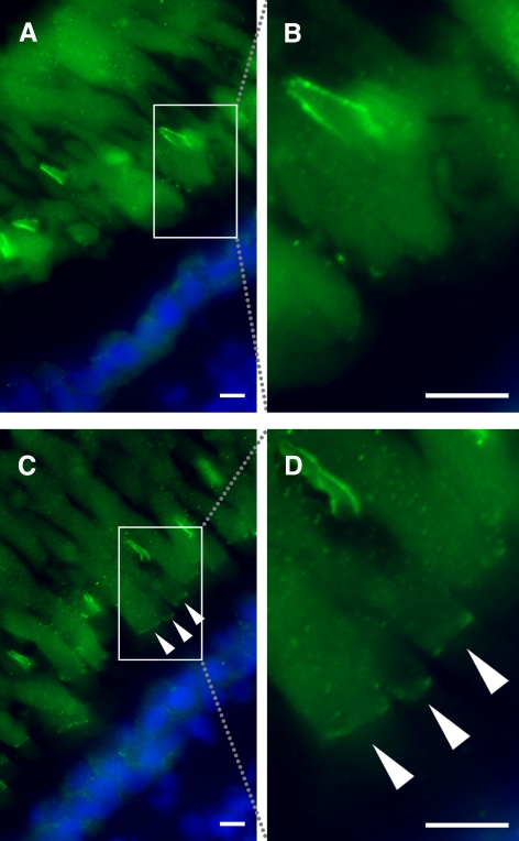
Variation in immunolabeling of X. laevis rods with anti–xlProminin-1 N terminus antibody αPN. (A) Immunolabeling of a retina from a tadpole euthanized at 4 hours before light onset with αPN (green). No labeling is seen at the bases of the ROS. The boxed area is enlarged as shown in (B). (C) Immunolabeling of a retina from a tadpole euthanized at 8 hours after light onset with αPN. Bases of the ROS are clearly labeled (arrowheads). The boxed area is enlarged as shown in (D). Nuclei are labeled with Hoechst 33342 dye (blue). Scale bars, 5 μm.

