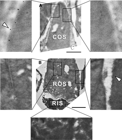
Figure 8.
Localization of xlProminin-1 with anti–xlProminin-1 C terminus antibody αPC in X. laevis photoreceptors examined by immuno-electron microscopy of frozen sucrose embedded retinas. (A) The rims of the disk lamellae of a COS opposite the side of the connecting cilium are labeled with αPC, detected with protein A 6 nm gold (arrowhead). Rims of the disk lamellae on the same side of the connecting cilium are not labeled. (B) Longitudinal section of a rod. ROS and RIS are not labeled with αPC, in contrast to an adjacent labeled COS membrane (arrowhead). Boxed areas are enlarged fourfold to show the details of typical immunolabeling of the sections. C, connecting cilium. Scale bars, 1 μm.

