Figure 9.
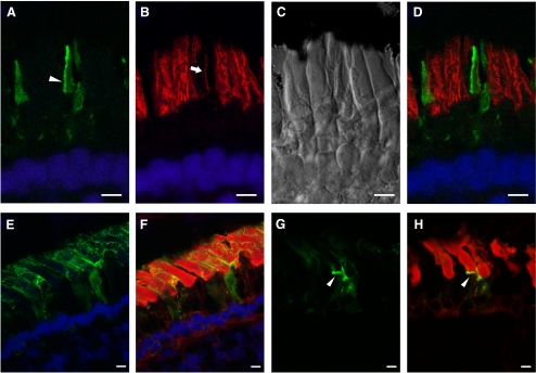
(A–D) xlProminin-1-hrGFP II expressed in transgenic X. laevis cones. Expression of the transgene is controlled by a newly isolated promoter XtCAP 1.9 for the X. tropicalis cone arrestin gene (see Supplementary Materials, http://www.iovs.org/lookup/suppl/doi:10.1167/iovs.11-8635/-/DCSupplemental). (A) The hrGFP II-tagged fusion protein localizes primarily to one side of COS (arrowhead). (B) The same section colabeled with anti–peripherin-2/rds antibody Xper5A11 (red). COS was labeled with the antibody and the labeling is confined to a thin area along one side of the COS (arrow). Longitudinal stripes in the adjacent rods result from the peripherin-2/rds localization along the aligned incisures of ROS disks. (C) Nomarski view of the same retina. (D) Superimposed image shows that xlProminin-1-hrGFP II resides on the side of the disk rims opposite peripherin-2/rds. Thus the transgenic hrGFP II-tagged protein expressed in cones distributes to the same sites as the endogenous protein we observed with anti–xlProminin-1 antibodies αPN and αPC. Nuclei are labeled with Hoechst 33342 dye (blue). (E–H) xlProminin-1-hrGFP II expressed in transgenic X. laevis rods. Expression of the transgene is controlled by a promoter XOP 1.3 for the X. laevis opsin gene. (E, F) The fusion protein is seen on the plasma membrane of both ROS and RIS (green) and occasionally is observed within the middle of the OS. Some fluorescence is seen within the RIS, presumably representing the trafficking of the newly synthesized protein. (G, H) The fusion protein is occasionally observed at the base of the ROS (arrowhead). Sections were counterstained with Texas Red-X conjugated wheat germ agglutinin (red) to visualize the ROS and post-Golgi membranes. Scale bars, 5 μm.

