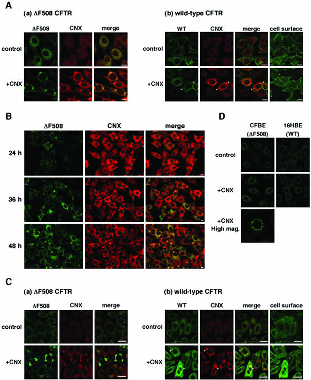
Figure 2.
Calnexin overexpression formed IB-like structures where ΔF508 CFTR and calnexin accumulated. (A) All panels are fluorescence micrographs of BHK cells stably expressing ΔF508 CFTR-GFP (a, ΔF508-BHK) and wt CFTR-GFP (b, CFTR-BHK). The cells were infected with or without Ad-CNX at a MOI 50 (+CNX), and 48 h after infection cells were fixed and permeabilized, immunostained with anti-calnexin antibody, and visualized with TRITC-conjugated secondary antibody. Analysis was performed by confocal laser scanning microscopy. GFP fluorescence is shown in the left panels (a, ΔF508 CFTR; b, wt CFTR), TRITC fluorescence in the middle panels (CNX), and right panels show the overlay (merge). Note that calnexin overexpression formed IB-like structures and ΔF508 CFTR specifically accumulated in the structures. Arrowheads represent the regions in which wt CFTR slightly accumulated with calnexin. (B) Time course of IB-like structure formation. ΔF508-BHK cells were infected with Ad-CNX (MOI 50) and incubated for the times indicated. Note that formation of IB-like structures of ΔF508 CFTR coincides with that of calnexin. (C) ΔF508-(a) and CFTR-CHO (b) cells were infected with or without Ad-CNX (MOI 50) and 48 h after infection cells were fixed and permeabilized, immunostained with anti-CFTR and anti-calnexin antibodies, and visualized with fluorescein isothiocyanate- and TRITC-conjugated secondary antibodies. (D) CFBE41o- cells (ΔF508 CFTR) and 16HBE14o- (wt CFTR) were infected with or without Ad-CNX (MOI 50), and 48 h after infection cells were fixed and permeabilized, immunostained with anti-CFTR antibody, and visualized with Alexa Flour488-conjugated secondary antibody. The cells were imaged so that the ER region was in focus, except for some images that were focused on the cell surface (cell surface). Bars, 10 μm.

