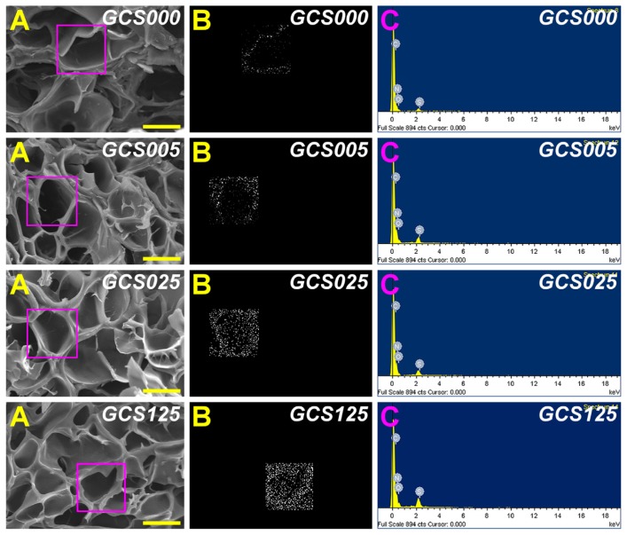
Figure 1.
Scanning electron microscopy/energy dispersive X-ray spectroscopy (EDS) mapping of various porous gelatin scaffolds modified with chondroitin-4-sulfate (GCS000, GCS005, GCS025, and GCS125): (A) scanning electron microscopy images, (B) EDS mapping of sulfur, and (C) EDS spectrogram.
Notes: Scale bars are 50 μm; scaffold groups labeled according to chondroitin-4-sulfate concentration used (0%, 0.05%, 0.25%, or 1.25% (w/v)): GCS000, GCS005, GCS025, and GCS125.

