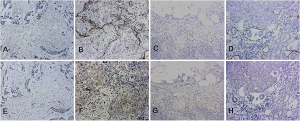
Figure 4.

Representative examples of D2-40 and ECM1 staining in the matched sections (100×). Sections with ECM1-positive staining had higher lymphatic microvessel densities. A-D: Lymphatic microvessels labelled with D2-40. E-H: ECM1 IHC staining: Panels E and G: ECM1 negative; Panels F and H: ++ ECM1 staining. A-B, E-F: invasive ductal breast cancer; C-D, G-H: lymph nodes with metastasis. Panels A and E, B and F, C and G, D and H represent matched sections obtained from the same specimen, respectively.
