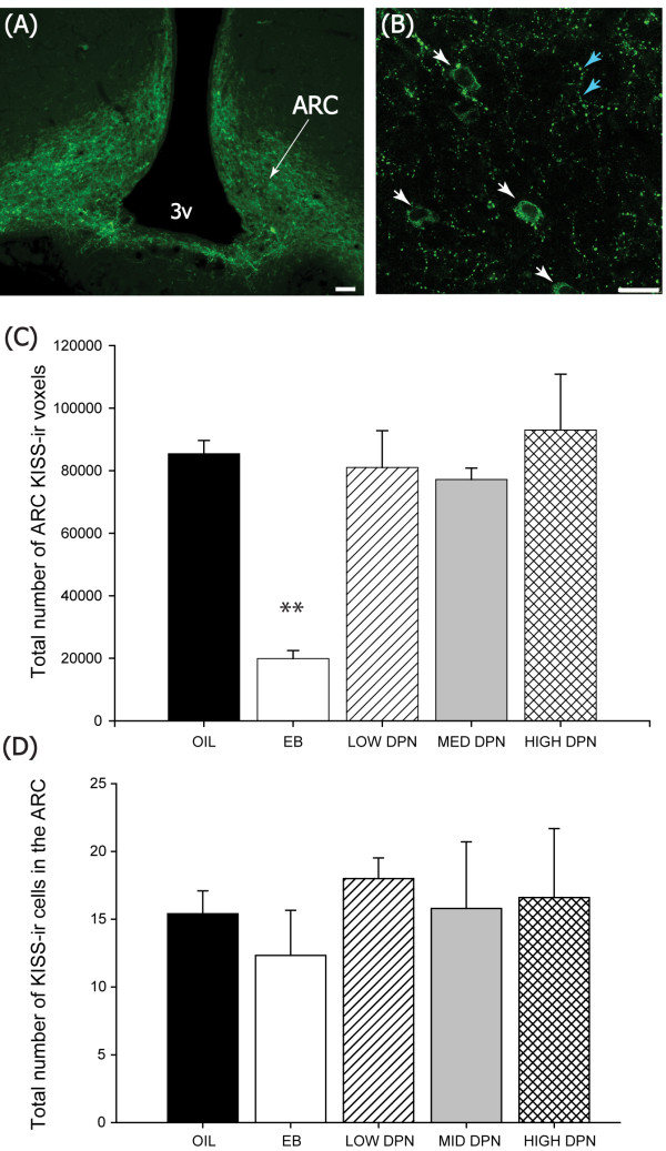
Figure 4.
Kisspeptin immunolabeling in the mediobasal hypothalamus. Fluorescence photomicrographs showing Kiss immunoreactive (-ir) labeling in the ARC of the hormone primed females (Experiment 1). Kiss immunolabeling was considerably denser (A) than in the AVPV (see Figure 3) and localized to both fibers, (B blue arrows) as well as a small number of cell bodies that were often difficult to discern from the heavy fiber labeling (B, white arrows). (C) The density of Kiss-ir labeling was significantly lower in the females neonatally treated with EB but not DPN at any dose examined. (D) The number of Kiss-ir cell bodies did not statistically differ between groups. (3v = 3rd ventricle, (A) Scale bar = 50 μm, (B) Scale bar = 25 μm, Means ± S.E.M., **P ≤ 0.001).

