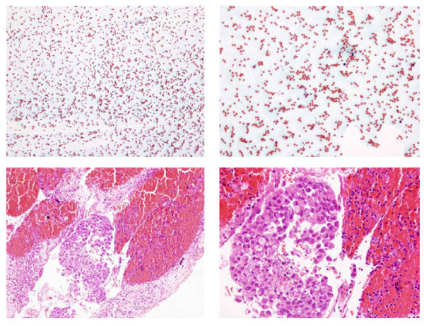
Figure 2.
Fine needle aspiration from a subcarinal node in a case of metastatic adenocarcinoma. Non-diagnostic conventional smear (figure 2a and b) and cluster of adenocarcinoma cells in the the cell block (figure 2c and d). a: Unsatisfactory specimen. Blood cells in an otherwise acellular smear (Papanicolau stain, x 100). b: Unsatisfactory specimen. Blood cells in an otherwise acellular smear (Papanicolau stain, x 200). c: Cluster of adenocarcinoma cells in a cell block (Hematoxilin and eosin stain, x 100). d: Cluster of adenocarcinoma cells in a cell block (Hematoxilin and eosin stain, x 200).

