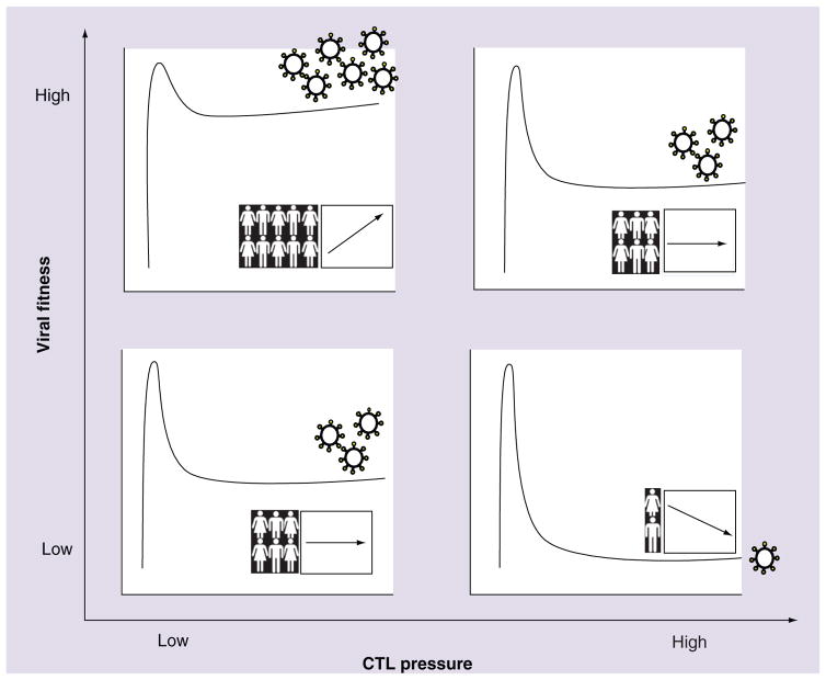
Figure 1. Individual and population-level implications of immune-mediated attenuation of HIV-1.
The extent to which immune-mediated fitness defects influence the clinical course of HIV infection, and the potential implications of immune-mediated HIV attenuation to the future of the epidemic, depend on various factors including the relative fitness of the variant acquired at transmission (y-axis) as well as the effectiveness of the host CTL response (x-axis). We present four simplified, hypothetical scenarios illustrating these combined effects on outcome for the individual (illustrated here as the kinetic curves of acute-phase HIV viremia and subsequent set point stabilization) and for the population (graph insets illustrating hypothetical trends in HIV incidence [human figures] and pathogenesis [arrows] over time). Upper-left quadrant: high transmitted viral fitness combined with poor host CTL responses yields high viral load set points in infected individuals. Upper-left quadrant inset: continued transmission of highly fit variants in the absence of immune selection pressure could result in increasing HIV pathogenesis (and potentially incidence) over the epidemic’s course. Lower-right quadrant: low transmitted viral fitness combined with highly effective host CTL responses yields successful immune control of HIV-1. Lower-right quadrant inset: continued passage of attenuated variants through hosts capable of mounting highly effective natural (or vaccine-induced) immune responses could result in decreasing HIV pathogenesis (and potentially decreased transmission risk) over time. Upper-right quadrant (and lower-left quadrant), plus insets: intermediate average plasma viral loads and stable population-level outcomes are envisioned in cases of high transmitted viral fitness and effective CTL responses (and vice versa).
CTL: Cytotoxic T lymphocyte.

