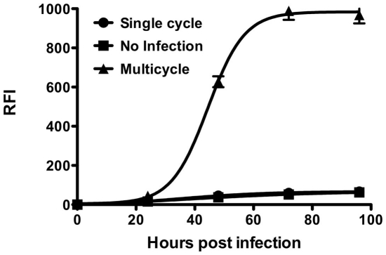
Figure 2. NiV multicycle replication using modified assay.
Cells were transfected with plasmids encoding NiV G and F (triangles) and then infected with pseudotyped VSV. Transfected cells were also left uninfected as a control (squares). Additionally, control cells transfected with empty plasmid were also infected with pseudotyped VSV, showing single cycle replication (circles). Relative fluorescent intensities of the RFP were measured after 24, 48, 72 and 96 hours.

