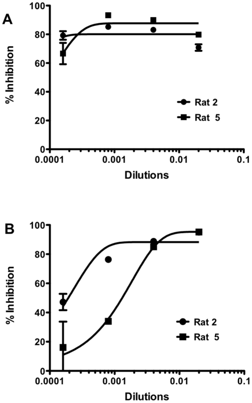
Figure 5. Validation of modified pseudotype assay validation for NiV.
Cells were transfected with plasmids encoding the NiV glycoproteins F and G and then infected with either pseudotyped NiV (A) or pseudotyped VSV (B) in presence of rat anti-NiV antibodies, Rat 2 (circles) or Rat 5 (squares). Relative fluorescent intensities of the RFP were measured after 48 hrs.

