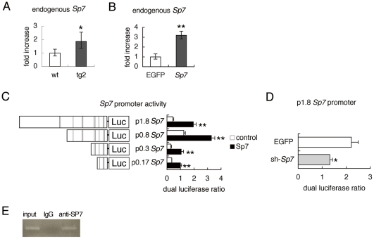
Figure 7. Positive regulation of Sp7 promoter by SP7.
(A, B) Real-time RT-PCR analysis. A, Endogenous Sp7 expression in wild-type and tg2 mice at 18 weeks of age. RNA was prepared from tibiae and femurs, in which hematopoietic cells were flushed out. *P<0.05 vs. wild-type mice. n = 4–5. B, Osteoprogenitors isolated from wild-type calvaria using collagen gel were infected with adenovirus expressing EGFP or Sp7 at 6×10⁶ pfu/well, and endogenous Sp7 expression was examined. **p<0.001 vs. EGFP-expressing cells. The level in wild-type mice or EGFP-expressing cells was set at 1, and relative values are shown. (C, D) Reporter assays using the Sp7 promoter. C, The reporter activity using the 1.8-kb Sp7 promoter-luciferase (Luc) construct and the deletion constructs. Reporter assays were performed using 293T cells with (closed columns) or without (open columns) the Sp7 expression vector. The lines in the schematic diagram of the Sp7 promoter indicate putative SP1-binding sites. D, The reporter activity using the 1.8-kb Sp7 promoter in ATDC5 cells infected with adenovirus expressing EGFP or sh-Sp7 at 6×10⁶ pfu/well. *p<0.05 and **p<0.001 vs. control. (E) ChIP assay. DNA before immunoprecipitation (input) and after immunoprecipitation with anti-SP7 antibody or anti-IgG antibody was amplified by PCR using primers that amplify the region containing the proximal 170 bp of Sp7 promoter.

