
Figure 4. Pharyngeal arch artery organization visualized by ink injection at E10.5.
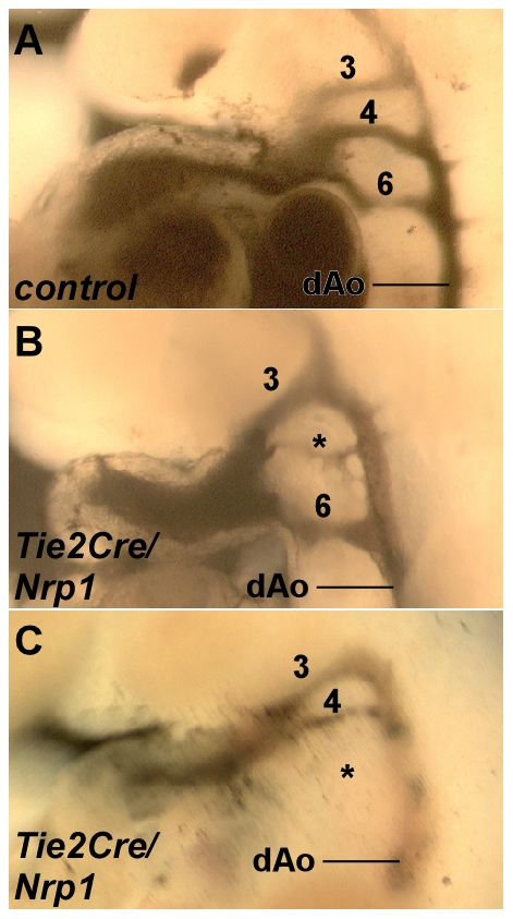
A, Normal arterial pattern in a control embryo; the numbers 3, 4, and 6 indicate the respective arch arteries. B,C, Examples of arch artery abnormalities seen in mutant embryos: an extremely hypoplastic 4th arch artery (B), and a missing 6th arch artery (C), both indicated by asterisks.