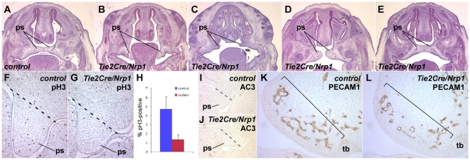
Figure 7. Reduced palatal mesenchyme cell proliferation in Tie2Cre/Nrp1 mutants.
A–E, frontal sections at E14.5 of control (A) and of several mutant embryos (B–E) showing a range of palatal phenotypes: failure of the palatal shelves (ps) to elevate (B), gross and mild failure to properly extend (C and D, respectively), and normal fusion (E; approx. half of mutants had normally fused palates). F,G, Proliferation detected by phospho-histone H3 immunohistochemistry in frontal sections of control (F) and Tie2Cre/Nrp1 (G) embryos at E13.5 (nuclei are counterstained with hematoxylin); the dotted line shows the approximate upper end of the palatal shelves for purposes of quantification of proliferating cells. H, Quantification of mesenchymal proliferation in E13.5 palatal shelves. I,J, Absence of palatal shelf apoptosis at E13.5 in frontal sections, visualized by immunostaining for activated caspase 3. K,L, Immunohistochemical detection of PECAM1 in control (K) and mutant (L) embryos (frontal sections) in the palatal shelves at E12.5. The bracket indicates the region of growth and extension; there are fewer and less well elaborated vessels in this region in the mutant. The avascular region behind the brackets in both panels is condensed mesenchyme that will form the maxillae. tb, tooth bud.

