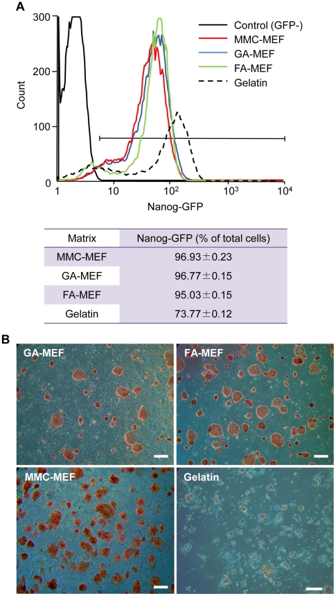
Figure 3. Maintenance of the undifferentiated state of miPS cells on MMC-treated MEFs or chemically fixed MEFs.
(A) Percentage of Nanog-GFP-expressing miPS cells cultured on different matrices. A gelatin-coated surface was used as a control matrix, as it is not able to maintain miPS cells fully in an undifferentiated state. EB3 cells were used as a negative control, as they have no GFP fluorescence. The expression of Nanog was analyzed using a fluorescence-activated cell sorter (FACS) after 10 passages on MMC-MEFs, six passages on GA-MEFs or FA-MEFs, and one passage on a gelatinized surface. (B) Alkaline phosphatase (AP) activity of miPS cells cultured on chemically fixed MEFs. Histochemistry revealed AP activity (red) in miPS cells cultured on GA-MEFs or FA-MEFs after six passages. Scale bars, 500 µm.

