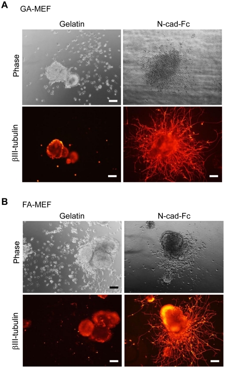
Figure 5.
Neural differentiation of miPS cells cultured on FA-MEFs (A) or GA-MEFs (B). After six passages on FA-MEFs or GA-MEFs, miPS cells were collected and neural differentiation was induced using the SFEB method in KSR medium supplemented with 5 µM SB43152 n-hydrate and 5 µM CKI-7. After 5 days of suspension culture, cells were then transferred to different matrices. βIII-tubulin immunostaining was performed to detect the formation of neural cells. Scale bars, 200 µm.

