
Figure 3.
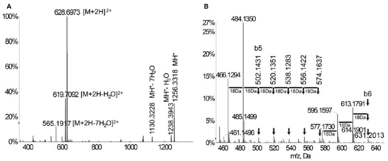
Ionization, fragmentation pattern, and serial neutral loss in primary and CID MS/MS of synthetic tubulin-α1A/1B CTT (EGEGEEEGEEY). (A) ESI-MS spectrum showing the singly charged synthetic CTT ion at m/z 1256.3318 and the double charged ion [M+2H]2+ at m/z 628.6973. Neutral losses of 1 water molecule were observed in both single-charged [M+H-H2O]+ (m/z 1238.3943) and double-charged [M+2H-H2O]2+ (m/z 619.7092) ions. Neutral losses of up to 7 water molecules were also detected for both ions: [M+2H-7H2O]2+ (m/z 565.1917); [M+H-7H2O]+ (m/z 1130.3228). (B) A close-up look at the CID MS/MS spectra of EGEGEEEGEEY showed 3x18Da neutral losses for b6 (m/z 631.2013) and 2x18Da loss for b5 fragments (m/z 502.1431) and a pattern of 7 peaks from m/z 466.1294 to m/z 574.1637 with a ΔM of 18Da.