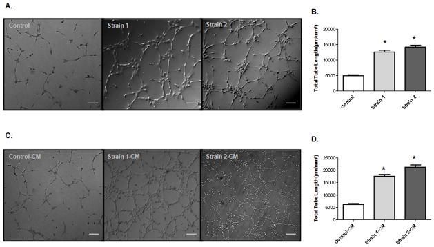
Figure 4.
PGI2-EPC strains promote in vitro angiogenesis. (A) Representative images of tubes formed in native EPCs (control) and PGI2-EPCs (strains 1 and 2) after being seeded on a reduced growth factor–membrane matrix. Scale bar=50 μm. (B) Quantification of tube formation. Total tube length was measured as μm/mm2, n=3; *P<0.05 vs control. (C) Representative images of tube formation in native EPCs in the presence of conditioned medium (CM) from native EPCs (control CM) and PGI2-EPCs (strain 1 and 2 CM). Scale bar=50 μm. (D) Quantification of tube formation. Total tube length was measured as μm/mm2, n=3; *P<0.05 vs control-CM.

