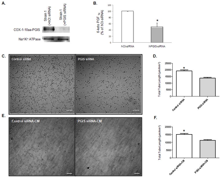
Figure 5.
Knockdown of hybrid enzyme expression in PGI2-EPCs decreased in vitro angiogenesis. (A) Western blot showed a significant reduction of COX-1-10aa-PGIS fusion protein in PGIS-EPC strain 1 after silencing of the human PGIS gene. Na+/K+ ATPase protein served as a loading control. (B) siRNA knockdown of human PGIS reduced prostacyclin production in the supernatant of PGI2-EPC strain 1. *P<0.05 compared to PGIS siRNA; n=3. (C) Representative images of tubular structures. Scale bar=50 μm. (D) Quantification of tube formation. Tube formation assays were performed 96 hours after transfection of human PGIS siRNA or control siRNA transfection into PGI2-EPCs. Total tube length was measured as μm/mm2, n=3; *P<0.05 compared to PGIS siRNA. (E) Representative images of tubular structures of native EPCs in the presence of conditioned medium (CM), which was collected 96 hours after PGIS siRNA or control siRNA transfection, respectively. Scale bar=50 μm. (F) Quantification of tube formation. Total tube length was measured as μm/mm2, n=3; *P<0.05 compared to PGIS siRNA-CM.

