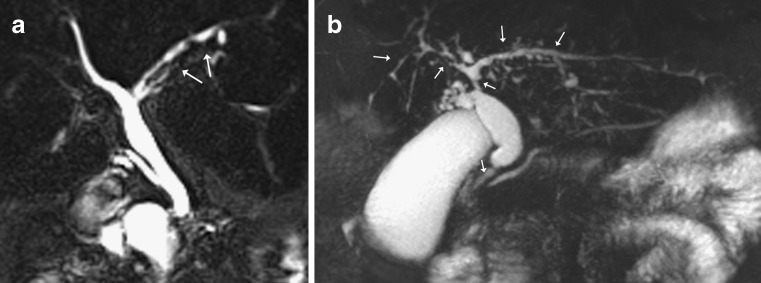
Fig. 10.
a Coronal MIP reformat shows early primary sclerosing cholangitis (PSC) with irregular dilatation and strictures seen in the left sided intrahepatic ducts (arrows). b Coronal MIP reformat in a different patient with more advanced PSC with multiple intrahepatic strictures and strictures seen in the common hepatic duct and distal CBD (arrows). There is dilatation of the proximal CBD

