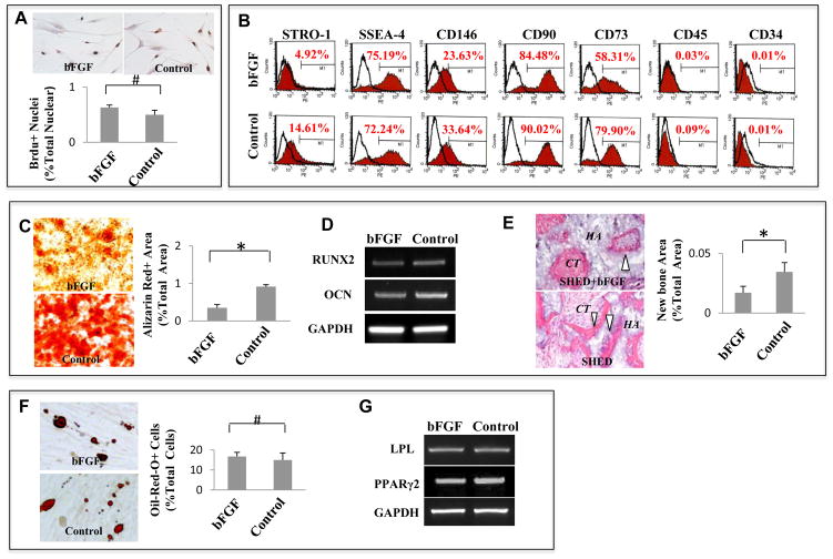
Figure 1. bFGF inhibits SHED osteogenic differentiation.
(A) bFGF treatment did not alter the proliferation rate of SHED as assessed by BrdU incorporation assay. The proliferation rate indicated as a percentage of BrdU+ nuclei to the total nuclear cells and averaged from five independent assays. (B) Flow cytometry showed that bFGF treatment reduced expression levels of STRO-1, CD 73, CD 90, and CD 146, but had no significant effect on expression of SSEA-4. SHED are negative to CD 45 and CD 34 antibody staining. (C–E) After osteogenic inductive culture for 4 weeks, bFGF-treated SHED showed a decreased mineralized nodule formation than untreated control group as assessed by alizarin red staining. Alizarin red-positive (Alizarin Red+) area corresponding to total area was averaged from five independent groups (C). After 2 weeks osteogenic induction, expression levels of osteogenic genes Runx2 and osteocalcin (OCN) were reduced in SHED treated with bFGF compared to untreated control group as assessed by RT-PCR. GAPDH was used as an internal control (D). SHED regenerated de novo bone (B white triangle) and connective tissue (CT) around HA/TCP carrier (HA) when transplanted subcutaneously into immunocompromised mice. bFGF-treated SHED formed less bone than untreated SHED. Newly formed bone area was calculated as a percentage of total area and averaged from three independent transplant assays (E). (F, G) Oil-red O staining showed that lipid accumulation in bFGF-treated SHED was similar to untreated SHED at 4 weeks adipogenic induction. Number of oil-red O-positive (Oil-Red-O+) cells was calculated as a percentage to total cells and averaged from five independent cultures (F). RT-PCR analysis indicated that expression levels of adipocyte-specific molecules LPL and PPARγ2 were no difference between bFGF-treated SHED and untreated SHED at 4 weeks post adipogenic culture. GAPDH was used as an internal control (G). (*P<0.05)

