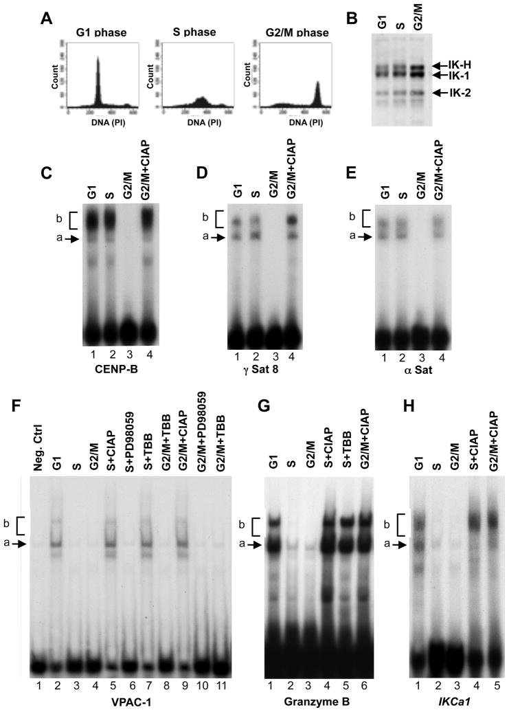
Figure 2. DNA binding of Ikaros during cell cycle.
(A) MOLT-4 cells were arrested in G1, in S, or in G2/M phase of the cell cycle, and assessed for DNA content by flow cytometry. (B) Nuclear extracts were obtained from cells at each phase, and normalized for Ikaros content by Western blot. (C–H) DNA binding of nuclear extracts from cells at indicated stages of cell cycle with probes derived from PC-HC (B–D) or the URE of indicated genes (E–G). Ikaros DNA-binding complexes are indicated as a) arrows indicating dimers that contain Ikaros isoforms and b) brackets indicating tetramers/higher order complexes of Ikaros isoforms.

