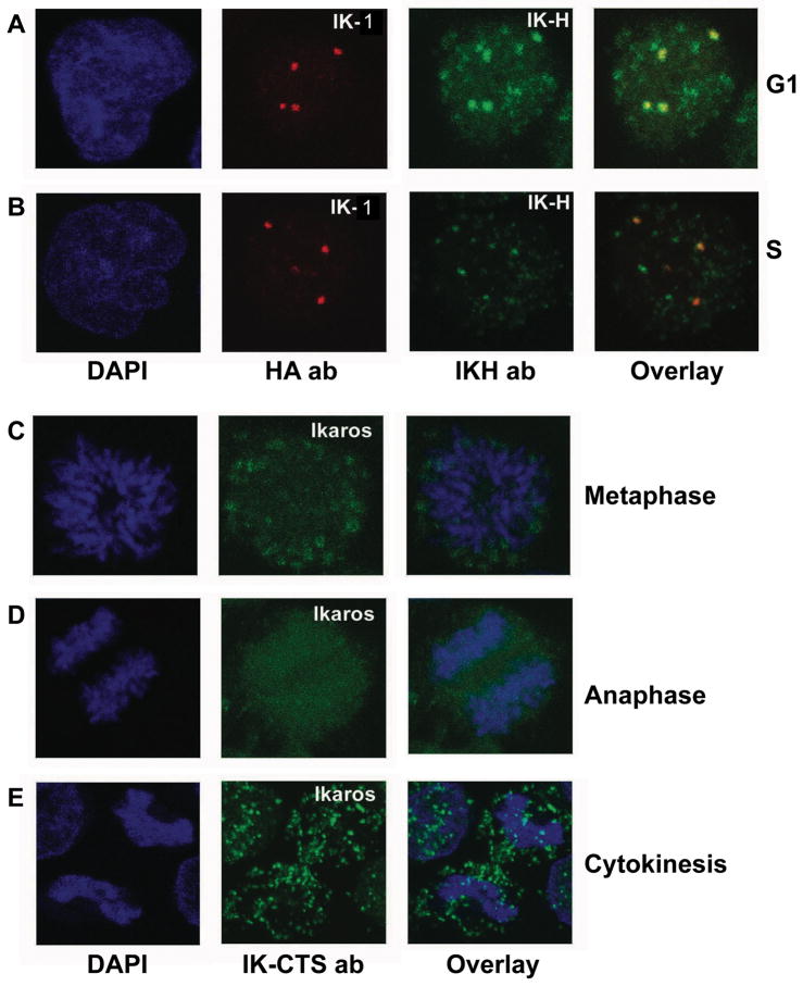
Figure 4. Subcellular localization of human Ikaros isoforms during cell cycle.
(A–B) CEM T-ALL cells infected with amphotrophic retrovirus expressing HA-tagged hIK-VI. Cells were arrested in G1 or S phase as described previously and stained for confocal microscopy. DAPI stain shows nuclear DNA (left panels). HA-IK-VI was detected with anti-HA antibody (red) and endogenous hIK-H with IK-H antibody (green). The combined image from anti-HA and IK-H staining is shown in the right panel. (C–E) Cycling CEM cells at various stages of cell cycle were selected based on DAPI staining (blue). Staining with IK-CTS antibody (green) detects all Ikaros isoforms. The combined image from DAPI and IK-CTS staining is shown in the right panel. Data shown are representative of cells at each stage.

