Figure 3.
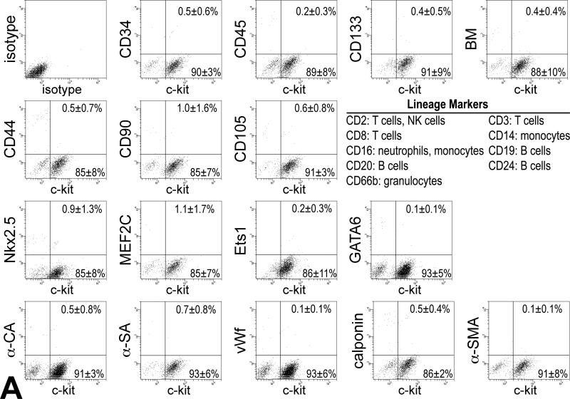
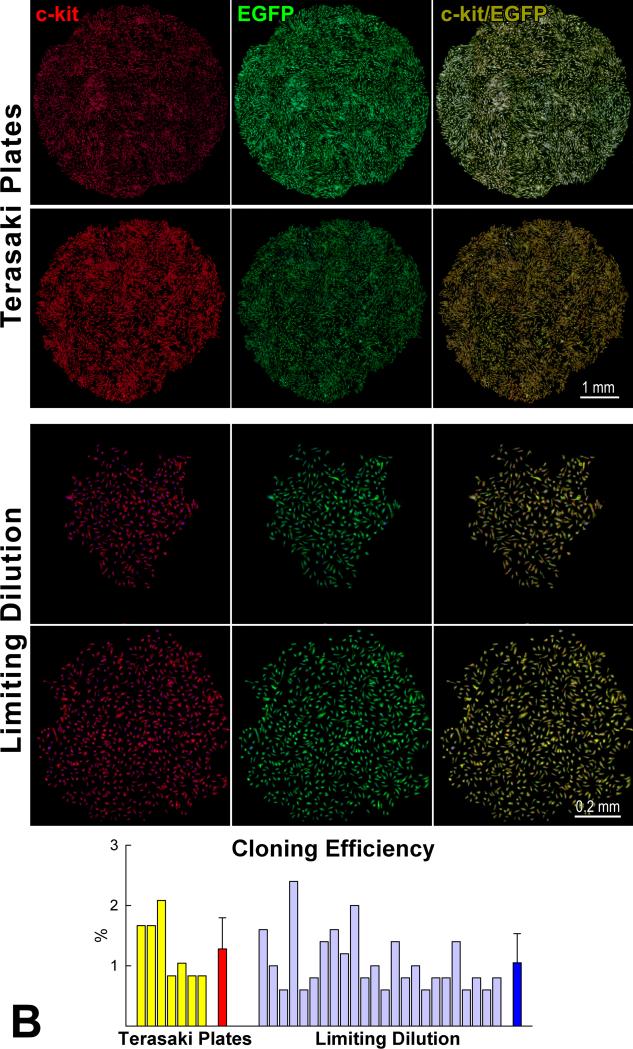
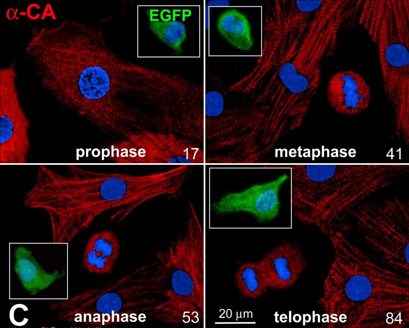
Characteristics of pCSCs. A: pCSCs were predominantly negative for markers of bone marrow cells, mesenchymal stromal cells, cardiomyocytes, ECs and SMCs. B: Four single cell-derived clones are shown (Terasaki plates: upper two; limiting dilution: lower two). Cells in the clones expressed c-kit (left, red) and EGFP (central, green). Right panel, merge. Cloning efficiency in each experiment is shown together with mean±SD. C: Dividing fetal myocytes in culture do not express EGFP. The number in each panel reflects sequential sampling of 102 cells. EGFP-positive CSCs (insets, green), positive control. α-CA, red. Chromosomes are stained by propidium iodide (blue).


