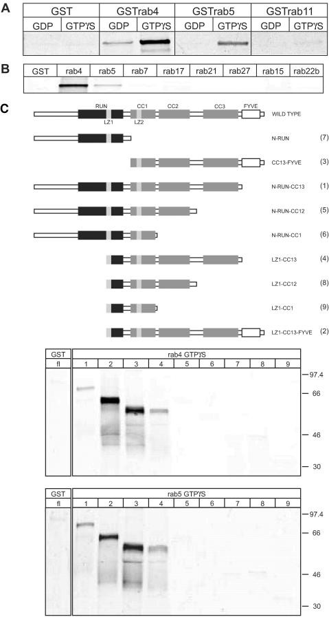
Figure 6.
rab4 and rab5 bind directly to rabip4′. (A) GST, GSTrab4, GSTrab5, and GSTrab11 were loaded with GTPγS or GDP and incubated overnight with35S-labeled VSV-G-tagged rabip4′. Beads were washed three times, and proteins were eluted with 25 mM reduced glutathione. Eluates were resolved on 10% SDS-PAA gels and analyzed by phosphorimaging. (B) GST, GSTrab4, GSTrab5, GSTrab7, GSTrab15, GSTrab17, GSTrab21, GSTrab22b, and GSTrab27 were loaded with GTPγS and incubated with 35S-labeled VSV-G-tagged rabip4′ as described in A. rabip4′ was eluted with glutathione and analyzed by SDS-PAGE and phosphorimaging. (C) Schematic representation of rabip4′ truncation mutants. See legend to Figure 1B for domain nomenclature. GSTrab4 and GSTrab5 were loaded with GTPγS. Binding and analysis of 35S-labeled VSV-G-tagged rabip4′ (fl) and truncations N-RUN-CC13 (1), LZ1-CC13-FYVE (2), CC13-FYVE (3), LZ1-CC13 (4), NRUN-CC12 (5), N-RUN-CC1 (6), N-RUN (7), LZ1-CC12 (8), and LZ1-CC1 (9) was done as described in MATERIALS AND METHODS.

