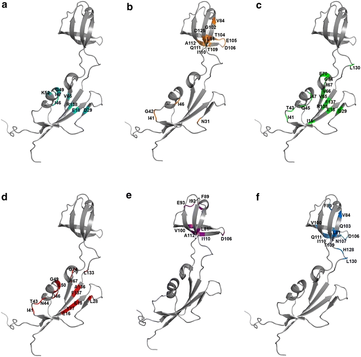
Fig. 5.

Structural mapping of the residues experiencing relatively large chemical shift perturbations upon interactions of the protein with a Ni2+, b twin-arginine translocation (Tat) signal peptide of HydA from H. pylori, c FK506, d rapamycin, e reduced and carboxymethylated α-lactalbumin, and f insulin. All experiments were conducted using HpSlyDΔC, except for Ni2+ titration, in which full-length HpSlyD was used
