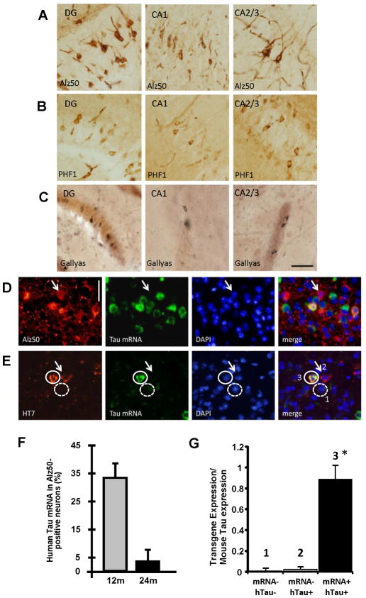
Figure 3. Tau propagates through neural circuits to mRNA-negative cells.
Several markers of pathological tau accumulation: Alz50 (A), PHF1 (B), and Gallyas silver staining (C), appear in brain regions synaptically connected to EC via the perforant pathway including DG, CA1, and CA2/3 (Scale bar 50 μm). FISH for tau mRNA (green), coupled with immunolabeling (red) with Alz50 (D) or HT7 (E) shows absence of mRNA in some cells containing the tau protein (arrows). Scale bar, 20 μm. Co-localization of tau mRNA and Alz50-positive aggregates was assessed by stereology at different ages, and compared to the total population of neurons carrying Alz50 aggregates. At 12 months of age, a third of the neurons affected by the tau pathology were expressing the transgene, while only ~3% were positive for MAPT gene at 24 months of age (F). FISH for MAPT mRNA (green), coupled with immunolabeling using HT7 (red) in the same EC sections from 17 month-old animals (E) was used to laser capture three different population of cells (G): 1) tau mRNA negative and human protein negative neurons; 2) mRNA negative and human tau protein positive neurons; 3) transgene mRNA positive and human tau protein positive neurons. (G) Total RNA was extracted and qPCR analysis of the cDNA product was carried out using primers against the transgenic human tau construct showing that the neurons which were human tau protein positive and were RNA negative by FISH did not express the tau transgene, confirming tau transmission to neurons that do not express the human tau transgene. Results are expressed as mean ± s.e.m. *P<0.001.

